In 1986, I spent my freshman year of college at a rolling suburban campus in St Louis, Missouri. Washington University, today ranked as one of the top five undergraduate business schools in the US and boasting a Pulitzer and Nobel Prize-winning faculty, enticed me away from my East Coast upbringing because it had a stellar English department. During my one year on campus (alas, this urban girl beat a hasty retreat to a northeastern city school) I waved the colors red and green, cheered the bear mascot through a miserable football season, and joined and dropped out of a cliquey sorority.
As I crisscrossed campus through green lawns and red brick buildings I had no idea that 25 years later the university would be a powerhouse of research, leading the way in the arenas of medical, environmental and energy, innovation and entrepreneurial and plant science. With over 3,000 projects utilizing $548.7 million in funding Wash U’s researchers are creating breakthroughs that will improve both our planet and our experience on it.
One of the current research rock stars is Lihong Wang, professor of biomedical engineering. A self-professed tool-maker, Wang excels in creating innovative medical technology and is leading the way in the field of photoacoustic imaging technique, a non-invasive process that uses the conversion of light and sound to create functional (physiological markers such as oxygenation and blood flow), molecular (biomarkers including gene activation and expression) and structural imaging.
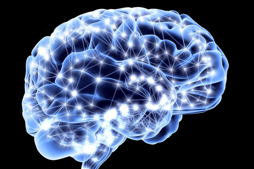
Inspired by President Obama’s BRAIN Initiative, Wang recognized that researchers could benefit from a tool he’d been perfecting. As he explains in an NPR interview, “We really don’t have a technique that allows us to see throughout the whole brain.” The problem: Functional MRI, PET scans and ultrasounds all have significant limitations. They offer slow and unclear images substantially restricted to structures close to the surface. Wang, however, has an idea for an alternative approach that will ultimately monitor brain activity in real time with an acuity fine enough to reveal an individual brain cell.
The crux of Wang’s stardom comes from his experiments with a process that combines the momentum and accuracy of light (which bounces around when it enters the body) and the penetrating ability of sound (which doesn’t bounce around). The result: photoacoustic imaging—and Wang—have exploded into the field of science and are bringing us ever closer to creating a full, 3D picture of the human brain. In fact, one of Wang’s research teams has already used a photoacoustic microscope to create such an image of a mouse brain. Using a laser to send pulses of light through a course of mirrors and filters, researchers sent the light into an anesthetized mouse brain. Although once inside the mouse’s skull the pulses of light bounce around enough of the light energy passes through to cause brain tissue molecules to vibrate; these vibrations produce sound waves that researchers captured in the form of an image. This is the essence of how photoacoustic imaging works: Biological tissues absorb light, which then converts to transient heating and subsequently ultrasonic waves that yield the image.
Wang’s research suggests numerous uses in brain science, yet the employment of photoacoustic imaging principles applies to other areas as well. Wang’s lab has developed processes that use photoacoustic imaging to reveal breast and skin tumors and even individual cancer cells in the blood. Even more broadly the use of photoacoustic imaging includes such preclinical processes as drug screening, biomarkers, gene activity and brain function in addition to clinical applications that span melanoma cancer screening, early response to chemotherapy, functional metabolism imaging and gastrointestinal tract endoscopy.
The potential for photoacoustic imaging makes it an attractive place for research dollars. While Wang as pioneer is the current “it” man in the field (he’s been lured away from Wash U to CalTech where his lab relocates next year) researchers around the world are pouring over problems, solutions and extended applications. In North America, Asia and Europe scientists are researching instrumentation, reconstruction imaging algorithms and probably also ways to breakthrough such photoacoustic imaging obstacles as limited light penetration in tissues and the inability of ultrasound signals to efficiently penetrate gas cavity or lung tissues.
While the details of photoacoustic imaging sound very 21st century hi-tech (indeed, its first application entered the field of biomedicine in the late 1980s), the process finds its roots in a much more low-tech time: the 1800s when legendary inventor, Alexander Graham Bell, first discovered that sound waves would form after light was absorbed by a material.
Over 100 years later scientists are finally realizing how to apply Bell’s discovery in ways that are inventive, diagnostic and also, perhaps, preventive if (or, perhaps, when) photoacoustic imaging makes the leap out of the lab and into the mainstream of medicine, healthcare and wellness.
Click here to get inspired by Rose’s easy steps to positively change your mind


